
| name | Amanita eremites | ||||||||
| author | Tulloss | ||||||||
| name status | nomen provisorum | ||||||||
| english name | "Hermit Lepidella" | ||||||||
| GenBank nos. |
Due to delays in data processing at GenBank, some accession numbers may lead to unreleased (pending) pages.
These pages will eventually be made live, so try again later.
| ||||||||
| intro |
Olive text indicates a specimen that has not been
thoroughly examined (for example, for microscopic details) and marks other places in the text
where data is missing or uncertain. The following material is based on original research of R. E. Tulloss. | ||||||||
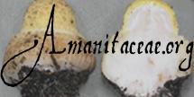
| pileus | 30 - 50 mm wide as dried, white to yellowish white as dried, hemispheric to convex; context pallid (probably white), rather thin compared to breadth of lamellae; margin nonstriate, appendiculate with subpulverulent-subfelted material and pieces of membranous partial veil, markedly incurved (at least at first), becoming rimose; universal veil as concolorous pulverulent squamules, densest over disc, possibly initially forming completely smooth surface layer. | ||||||||
| lamellae | free, crowded, cream with brown tint near edge as dried, quite broad, rounded at outer end, with minutely flocculose white edge; lamellulae rounded truncate to subattenuate to attenuate, plentiful, of diverse lengths. | ||||||||
| stipe | 23 - 43 × 3+ mm as dried, concolorous with pileus (as dried), approximately cylindric, longitudinally striatulate, decorated with white, flocculose squamules below partial veil; bulb 13 - 18 × 4 - 7 mm as dried, narrowly clavate to narrowly fusiform, often pointed below; context white in exsiccata; partial veil concolorous (probably white), superior, membranous, satiny as dried, thin, somewhat persistent, becoming radially lacerate; universal veil as squamules on stipe and as felted material appressed to bulb and/or in fine rings around stipe base above bulb or as short, slightly flaring, slightly lacerate, submembranous/felted limb. | ||||||||
| odor/taste | not recorded. | ||||||||
| macrochemical tests |
none recorded. | ||||||||
| pileipellis | absent; only a narrow and ungelatinized layer of hyphae found between the structures of the universal veil and the pileus context; filamentous undifferentiated hyphae with yellowish walls particularly dense in this layer and in lower universal veil and upper pileus context (overall, in region 600 - 700 µm wide). | ||||||||
| pileus context | filamentous undifferentiated hyphae 1.5 - 14.0 µm wide, branching, dominating, sometimes in fascicles, having both colorless and yellowish (subrefractive) walls, with many intercalary segments partially inflated (taking form of inflated cells of universal veil, up to 21 µm wide, with both colorless and yellowish walls); acrophysalides scattered, unevenly distributed, thin-walled, colorless, ovoid to ellipsoid to clavate to broadly fusiform, up to 88 × 46 µm; vascular hyphae not observed. | ||||||||
| lamella trama | bilateral; wcs = 70 - 90 µm; with angle of divergence from shallow to about 60°; wst-near = 45 - 90 (-95) µm; wst-far = 65 - 100 µm; with subhymenial base consisting of smoothly curving rather broad hyphae (3.0 - 14.0 µm wide, with many 10± µm wide) with constricted hyphae terminating at (and giving rise to) subhymenium; filamentous undifferentiated hyphae 1.5 - 14.0 µm wide, branching; diverging, terminal inflated cells not observed; vascular hyphae not observed. | ||||||||
| subhymenium | pseudoparenchymatous, 3 - 4 cell layers thick, with larger cells (e.g. 15.8 × 12.5 µm) arising from curving hyphae of subhymenial base, with cells generally decreasing in size approaching bases of basidia. | ||||||||
| basidia | 34 - 44 × 9.2 - 11.0 µm, 4-sterigmate, with sterigmata up to 8.0 × 3.0 µm; clamps common, prominent. | ||||||||
| universal veil | On pileus: with periclinal orientation, intergrading with pileus context (see under PILEIPELLIS, above); filamentous undifferentiated hyphae 1.8 - 8.8 µm wide, with both colorless and yellowish (subrefractive) walls, branching, with yellowish walled hyphae most common in region nearest pileus context, with yellowish walled hyphae sometimes loosely coiled and suggesting vascular hyphae; inflated cells elongate-ellipsoid to subventricose to narrowly clavate, to 99 × 26 µm, with walls thin or very slightly thickened; vascular hyphae not observed; clamps common, prominent, with those on yellowish walled hyphae concolorous.nbsp; On stipe and upper bulb: similar to that on pileus, but with much greater proportion of filamentous undifferentiated hyphae, with yellowish walled hyphae much less common than on pileus, with some gelatinization near surface; inflated cells up to 100 × 24 µm; clamps present, prominent. | ||||||||
| stipe context | longitudinally acrophysalidic; filamentous, undifferentiated hyphae 2.2 - 10.5 µm wide, plentiful to (locally) dominating, branching, densely packed, with those of largest diameter having walls up to 0.5 µm thick, sometimes with slightly inflated intercalary segments, with septa often constricted; acrophysalides barely inflated to narrowly clavate, up to 209 × 24 µm, with walls as in filamentous, undifferentiated hyphae (up to 0.8 µm thick), plentiful, unevenly distributed, locally clustered; vascular hyphae not observed; clamps present, prominent. | ||||||||
| partial veil | filamentous, undifferentiated hyphae 1.8 - 12.0 µm wide, dominant, dominantly subradially arranged, frequently branching, moderately loosely interwoven, often in fascicles, with yellowish walled hyphae as plentiful as in universal veil on pileus, with some inflated intercalary cells and slightly inflated tip cells; inflated cells narrowly clavate to narrowly fusiform, terminal, up to 88 × 15.0 µm; vascular hyphae not observed; clamps present, often prominent, concolorous with hyphae on which found (i.e., concolorous or yellowish-walled). | ||||||||
| lamella edge tissue | sterile. | ||||||||
| basidiospores | composite of data from all material revised by RET: [80/4/1] 9.2 - 11.2 (-12.0) × (7.2-) 7.8 - 9.0 (-10.2) µm, (L = 10.2 - 10.4 µm; L’ = 10.3 µm; W = 8.2 - 8.3 µm; W’ = 8.3 µm; Q = (1.03-) 1.15 - 1.34 (-1.39); Q = 1.24 - 1.25; Q’ = 1.25), hyaline, colorless, thin-walled, smooth, amyloid, subglobose to broadly ellipsoid, infrequently globose, occasionally ellipsoid, usually at least somewhat adaxially flattened; apiculus sublateral, stubby and truncate-conic, or slender and cylindric; contents monoguttulate with additional small granules; color in deposit unknown. | ||||||||
| ecology | In desert, within bounds of Desert Botanical Garden, growing with Chlorophyllum molybdites Mass. [Note: The collector responded to a letter from RET (11 May 1993) saying that he recalled collecting a fungus on about the date of this collection in an area in which seed received from all over the world was washed. He raised the possibility that the present species may have been imported from an unknown source. Viability of Amanita spores over an extended period of time has not been demonstrated. If the hypothesis is correct, living mycelium would possibly have been imported. Mr. Haughey also recalls that the plants nearest the collecting site were Larrea tridentata (Creosote Bush) and Cercidium microphyllum (Foothills Paloverde).—RET] | ||||||||
| material examined | U.S.A.: ARIZONA—Maricopa Co. - Phoenix, Desert Botanical Garden, ca. 90 m W of herbarium, 15.vii.1975 Russell Haughey s.n. (DES 16735, mixed collection labeled “Chlorophyllum molybdites”). | ||||||||
| discussion |
This species is only described provisionally because of the lack of notes on fresh material. DES 16735 consists of one mature basidiome of Chlorophyllum molybdites and five basidiomes of the present species. Four of these latter are mature with plentiful spores present on hymenial surfaces. The subhymenial tree of this species is very much like that illustrated for A. vittadinii by Bas (1969: fig. 31). | ||||||||
| citations | —R. E. Tulloss | ||||||||
| editors | RET | ||||||||
Information to support the viewer in reading the content of "technical" tabs can be found here.
Each spore data set is intended to comprise a set of measurements from a single specimen made by a single observer; and explanations prepared for this site talk about specimen-observer pairs associated with each data set. Combining more data into a single data set is non-optimal because it obscures observer differences (which may be valuable for instructional purposes, for example) and may obscure instances in which a single collection inadvertently contains a mixture of taxa.


Text and User-Generated Sporographs are published under the Creative Commons License.

In the case of a taxon page, image credits are on the 'image' tab.
